Ultrasound devices use sound waves with frequencies higher than the upper audible limit of human hearing. Ultrasound is no different from ‘normal’ (audible) sound in its physical properties, except in that humans cannot hear it. This limit varies from person to person and is approximately 20 kilohertz (20,000 hertz) in healthy, young adults. Ultrasound devices operate with frequencies from 20 kHz up to several gigahertz.
Some of ultrasound modes:
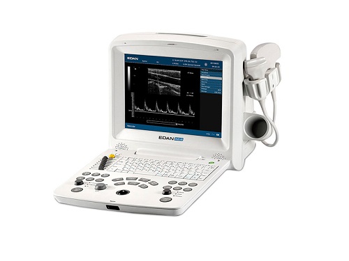
A-mode ultrasound
In ultrasonography a display in which imaging data are represented as echo amplitudes (on the y-axis) and time (on the x-axis), similar to the way electromagnetic waves are represented on an oscilloscope.
Synonym: A-mode; A-mode (amplitude modulation) display
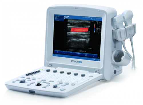
B-mode ultrasound
In ultrasonography, a display that uses dots of differing intensities to represent echoes received from tissues that more strongly or weakly reflect sound waves.
Synonym: ; B-scan
M-mode ultrasound
An ultrasonic display mode in which the motion of structures is seen on the vertical axis of the display, used, e.g., to show the movement of the heart’s valves and walls during diastole and systole.
Synonym: motion-mode display; time-motion mode ultrasound
continuous wave ultrasound
A form of ultrasound used in echocardiography in which a dual crystal transducer continuously generates and receives an ultrasound signal. It is used to measure blood velocities, e.g., across heart valves. A serious shortcoming of continuous wave ultrasound is its inability to identify depth accurately.
continuous wave Doppler ultrasound
Doppler ultrasonography that uses spectral Doppler in a constant series of echoes both originating and being received by the same transducer. It is used to study obstruction to blood flow through vessels.
duplex Doppler ultrasound
Doppler ultrasonography that uses a transducer with two functions: pulsed-wave Doppler and B-mode imaging.
endobronchial ultrasound
Abbreviation: EBUS
The use of ultrasonic transducers carried within a bronchoscope to evaluate tissues in or adjacent to the trachea and bronchi. EBUS can be used to identify solid masses to be biopsied. It helps distinguish solid masses, which may be malignant, from blood vessels such as the aorta or pulmonary arteries, which should not be penetrated with a biopsy needle.